Bone healing
When bone is fractured skeletal stem and progenitor cells activate, proliferate and differentiate into chondrocytes. These chondrocytes will form a cartilaginous template for new bone formation. As blood vessels enter this template, osteoprogenitors come along1, and differentiate into mineralizing osteoblasts. Now, that the template is mineralized, bone-resorbing osteoclasts mingle and reshape the template until the bone is perfectly shaped as before.
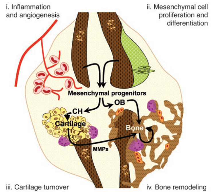
Understanding the process of fracture healing is of great importance, as 5-10 % of all fractures cannot heal properly. When this happens it severely impacts a person’s life quality, and increases the probability on mortality. As the process of bone formation after a fracture all starts with the skeletal stem cells that divide and give rise to the chondrocytes and osteoblasts, many researchers in the bone field are interested in how this activation process works, and if we could influence it, in order to repair non-healing fractures more easily. Researcher from KU Leuven and Harvard recently discovered how the inflow of nutrients affects the differentiation path the skeletal progenitor cells.3
Article: “Lipid availability determines fate of skeletal progenitor cells via SOX9”
In their paper, they described an increase in chondrogenesis when blood vessels are unable to enter the fractured region. Seven days after the fracture they noticed that blood vessels from the muscles were tightly interwoven with the fracture site and wondered how this interaction was established. Firstly, they examined if the periosteum cells, the cells laying the outer bone surface, contributed to the blood vessel formation. Although they observed that the progenitor cells in the periosteum did not give directly rise to blood vessels, when they removed the periosteum layer, the infiltration of blood vessels was inhibited, therefore suggesting that the periosteum actively promotes the ingrowth of blood vessels. Secondly, they wanted to study the effect on bone repair when blood vessels are absent. Therefore they inserted a filter, wherethrough blood vessels are unable to enter, between the site of injury and the muscle. This resulted in the increase of chondrocytes and reduced bone at the site of injury, together with the increased expression of Sox9, the main transcription factor to start chondrogenic differentiation.
As blood vessels supply the tissue with different types of nutrients, the researchers wanted to know which nutrients were crucial. By depriving cells in vitro with specific nutrients, such as oxygen, glucose and serum, they observed most pronounced effects on the Sox9 levels with the deprivation of serum, a main source for lipids. Moreover, addition of fatty acids to an ex vivo serum deprived culture restored the increased expression of Sox9, and in vivo injection of extra fatty acids into a fracture reduced the amount of chondrocytes present. This suggested that the lipids supplied by blood vessels during fracture healing determine chondrogenic differentiation of the progenitor cells.
Now, which cells need lipids in order to function normally? To answer this question, the researchers looked at the metabolism of osteoblasts and chondrocytes. By using single-cell RNA sequencing of mouse long bones, they found that chondrocytes highly rely on glucose metabolism, whereas osteoblasts use glucose and lipids to obtain their energy. This was confirmed by injecting fluorescent labeled fatty acid and glucose analogues. In this experiment they saw that whereas the glucose was taken up by both cell types, only the osteoblasts consumed the lipids. Therefore, lipids present or not, chondrocytes will survive and thrive anyhow.
Then they looked how the lipid scarcity affects the skeletal stem and progenitor cells. They saw that when skeletal progenitor cells were deprived from lipids, they first responded with the translocation of lipid droplets to the mitochondria, where fatty acid oxidation takes place. This way the skeletal progenitor cells still could maintain themselves within the first hours of lipid scarcity. Afterwards, levels of Sox9 rose concomitantly with the lipid deprivation, indicating a correlation. Therefore, the researchers deleted the Sox9 gene and looked at the response on the lipid metabolism. They saw that cell lacking Sox9 deprived from serum, the main source of lipids, continued lipid acid oxidation, whereas normal cells switched from lipid to glucose metabolism. On the contrary, overexpression of Sox9 reduced lipid consumption, which indicates that Sox9 is a metabolic regulator by suppressing lipid metabolism.
So how does the presence of lipids affects Sox9 levels, which in turn affects lipid metabolism? Upon serum deprivation, the researchers saw an enrichment of the FOXO/forkhead motif coinciding with localization of FOXO1 and FOXO3a protein to the nucleus, where it is transcriptionally active. Inhibition of FOXO signaling seemed to block the increase of Sox9 levels upon lipid deprivation, which indicates FOXO directly affects Sox9 expression during lipid deprivation.
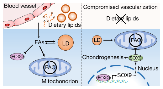
These results are of great importance, as they have potential clinical applications in the field of regenerative medicine, as altering the supply of nutrients or manipulating cellular metabolism might drive the progenitor cells into a desired differentiation pathway.
For more details about the experimental work:
Gastel, Nick Van, Stegen, Steve, Eelen, Guy, Schoors, Sandra, Baryawno, Ninib, Przybylski, Dariusz, Depypere, Maarten, Stiers, Pieter-jan, Lambrechts, Dennis, Looveren, Riet Van, et al. (2020). Lipid availability determines fate of skeletal progenitor cells via SOX9. Nature 579, 111–117
References:
1. Maes, Christa, Kobayashi, Tatsuya, Selig, Martin K., Torrekens, Sophie, Roth, Sanford I., Mackem, Susan, Carmeliet, Geert & Kronenberg, Henry M. (2010). Osteoblast Precursors, but Not Mature Osteoblasts, Move into Developing and Fractured Bones along with Invading Blood Vessels. Dev. Cell 19, 329–344
2. Maes, Christa, Coenegrachts, Lieve, Stockmans, Ingrid, Daci, Evis, Luttun, Aernout, Petryk, Anna, Gopalakrishnan, Rajaram, Moermans, Karen, Smets, Nico, Verfaillie, Catherine M., Carmeliet, Peter, Bouillon, Roger & Carmeliet, Geert. (2006). Placental growth factor mediates mesenchymal cell development , cartilage turnover , and bone remodeling during fracture repair. Jci 116, 16–18
3. Gastel, Nick Van, Stegen, Steve, Eelen, Guy, Schoors, Sandra, Baryawno, Ninib, Przybylski, Dariusz, Depypere, Maarten, Stiers, Pieter-jan, Lambrechts, Dennis, Looveren, Riet Van, et al. (2020). Lipid availability determines fate of skeletal progenitor cells via SOX9. Nature 579, 111–117
 Bone Biology & Beyond