My name is Dana Trompet, and I am a PhD student in the lab of Skeletal Cell Biology and Physiology at KU Leuven, Belgium. I am passionate about science and would like to share new interesting ideas within my field of expertise.
Osteoporosis is characterized by a decreased bone mass, leading to fragile bones more prone to fractures. This is caused by an imbalance in bone formation, when the bone-forming osteoblasts cannot catch up with the bone-resorbing osteoclasts. Previously, drugs used for the treatment of osteoporosis, such as bisphosphonates and denosumab, focused on inhibiting the bone resorption, which, when given intermittently, is effective for a period of time, however, after time this effect wears off and might even lead to serious side effects. Recently, two new osteo-anabolic drugs have been released into the marker, namely Romosozumab and Blosozumab, both monoclonal antibodies targeting sclerostin.
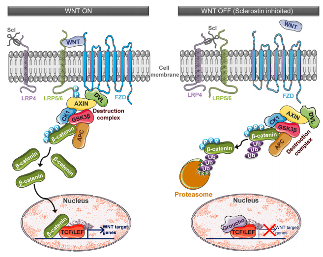
Sclerostin is an important regulator of the Wnt signaling pathway. When Wnt signaling is activated (‘WNT ON’), Wnt ligand binds to the low-density lipoprotein receptor-related protein 5/6 (LRP5/6) and FZD co-receptors, resulting in the intracellular phosphorylation of LRP5/6. This induces the inactivation of the destruction complex, which consists of dishevelled (DVL), AXIN, casein kinase 1 (CK1) and glycogen synthase kinase 3 beta (GSK3β), as it will localize to the cell membrane where its kinase activity is inhibited. This allows non-phosphorylated β-catenin to accumulate and translocate to the nucleus, where its transcriptionally active. Sclerostin (Scl), however, works as a Wnt signaling antagonist, as it binds to the LRP co-receptors, preventing their interaction with the Wnt ligands (‘WNT OFF’). This leads to the phosphorylation of β-catenin by the destruction complex, thereby labelling it for ubiquitination and proteasomal degradation.
Image taken from Holdsworth, Gill, Roberts, Scott J. and Ke, Hua Zhu (2019) ‘Novel actions of sclerostin on bone’, Journal of Molecular Endocrinology, 62(2), pp. R167–R185
As sclerostin normally prevents the activation of Wnt signaling, resulting in decreased bone formation capacity, the inhibition of sclerostin stimulates bone formation, making it a potential effective osteo-anabolic treatment.
Reference:
Holdsworth, Gill, Roberts, Scott J. and Ke, Hua Zhu (2019) ‘Novel actions of sclerostin on bone’, Journal of Molecular Endocrinology, 62(2), pp. R167–R185.
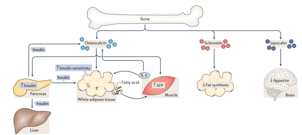
Bone has long been thought of to be a static organ, providing mechanical support to the body. However, recently several discoveries have shown that bone could also act as an endocrine organ, in which it signals to other organs, influencing glucose metabolism, energy expenditure and male fertility (Karsenty, 2011). More specifically, signaling between the bone and muscle has been described by studying the musculoskeletal system. This system can be seen as one complex organ, which comprises of skeletal bones, muscle, tendons, ligaments, cartilage and joints. These individual parts must be in efficient communication with one another to provide proper locomotion (Brotto and Johnson, 2014).
Image derived from (Dirckx et al., 2019); Dirckx, Naomi, Moorer, Megan C., Clemens, Thomas L. and Riddle, Ryan C. (2019) ‘The role of osteoblasts in energy homeostasis’, Nature Reviews Endocrinology. Springer US, 15(11), pp. 651–665.
Osteocalcin is a protein specifically secreted by the osteoblasts during bone formation and is therefore often used as a serum marker for bone formation. Post-translationally it undergoes carboxylation, which increases its affinity to the hydroxyapatite crystals, facilitating the embedding into the bone matrix. Upon bone resorption, which initiates with the formation of an acidic environment by osteoclasts, osteocalcin is decarboxylated and therefore loses its affinity to the bone matrix and promotes the entrance into the circulation (Mizokami, Kawakubo-Yasukochi and Hirata, 2017). In a recent publication, Subrata Chowdhury and colleagues describe the mechanism how osteocalcin secretion by osteoblasts enhances muscle function during endurance exercise (Chowdhury, 2020).
Title: “Muscle-derived interleukin 6 increases exercise capacity by signaling in osteoblasts”
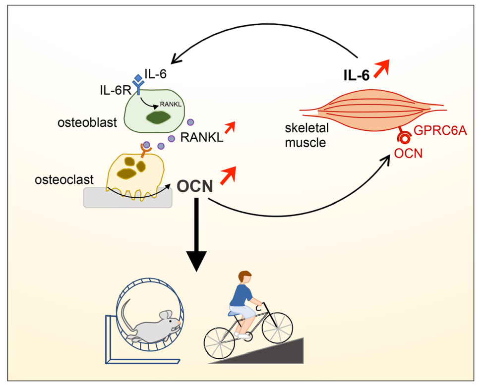
In this work, they studied the interplay between interleukin-6 and osteocalcin upon exercise. Firstly, they observed a correlation between osteocalcin and interleukin-6 (IL-6) serum levels; IL-6 levels increased upon osteocalcin injection in several mammals, and in humans both IL-6 and osteocalcin serum levels increased after exercise. Secondly, they conditionally deleted IL-6 from muscle myofibers by crossing the Hsa-MerCreMer mice with IL-6 floxed mice. In this model, no IL-6 was increased in the serum upon exercise, indicating that IL-6 is mainly muscle-derived. To determine if the changes in serum levels of IL-6 and osteocalcin where directly correlated, they generated heterozygote Ocn+/- IL-6+/- mice. These mice showed similar phenotype compared to the IL-6 conditionally deleted model, indicating a direct connection between the two proteins. An injection of osteocalcin could rescue the reduced exercise capacity, indicating that osteocalcin regulates the IL-6 serum levels.
To uncover an interplay between IL-6 and osteocalcin, the researchers were interested in the cell type present in the bone (osteoblast or osteoclast) that responded to the increased IL-6 levels. To study the interplay between osteoblasts and osteoclasts, they performed a co-culture, where both cell types are cultured within the same medium but aren’t physically touching. This allows the communication with one another by secreted signaling molecules, without any direct contact. They deleted IL-6 receptor in osteoblasts or osteoclasts prior to the co-culture, and studied the effects on osteoclastogenesis. They found that deletion of the IL-6 receptor in osteoblasts, not in osteoclasts, influenced the osteoclastic differentiation, indicating that IL-6 signals through osteoblasts and consequently affects the osteoblast-osteoclast coupling.
To validate this in vitro finding in vivo, they conditionally deleted IL-6 receptor in osteocalcin expressing osteoblasts. Strikingly, these mice showed similar reduction in endurance exercise as they had seen in the conditional model where they deleted muscle-derived IL-6. These mice did not show any increase in osteocalcin upon exercise and the observed phenotype could be partially rescued by administration of osteocalcin, whereas administration of IL-6 did not. Hence, this in vivo model proves that IL-6 signaling through osteoblasts is required for the beneficial osteocalcin derived effects on exercise.
In this study, the researchers demonstrate a new pathway in which bone signals to muscle upon exercise. This research widens the knowledge of the endocrine functions described to bone and makes the field wonder about a more general role of bone and bone-derived cells and their interaction and role within the whole body.
Image derived from Chowdhury S, Schulz L, Palmisano B, et al. Muscle-derived interleukin 6 increases exercise capacity by signaling in osteoblasts [published online ahead of print, 2020 Apr 27]. J Clin Invest. 2020;133572. doi:10.1172/JCI133572
References:
Chowdhury S, Schulz L, Palmisano B, et al. Muscle-derived interleukin 6 increases exercise capacity by signaling in osteoblasts [published online ahead of print, 2020 Apr 27]. J Clin Invest. 2020;133572. doi:10.1172/JCI133572
Brotto, Marco and Johnson, Mark L. (2014) ‘Endocrine crosstalk between muscle and bone’, Current Osteoporosis Reports, 12(2), pp. 135–141.
Dirckx, Naomi, Moorer, Megan C., Clemens, Thomas L. and Riddle, Ryan C. (2019) ‘The role of osteoblasts in energy homeostasis’, Nature Reviews Endocrinology. Springer US, 15(11), pp. 651–665.
Karsenty, Gerard (2011) ‘Bone endocrine regulation of energy metabolism and male reproduction’, Comptes Rendus – Biologies. Academie des sciences, 334(10), pp. 720–724.
Mizokami, Akiko, Kawakubo-Yasukochi, Tomoyo and Hirata, Masato (2017) ‘Osteocalcin and its endocrine functions’, Biochemical Pharmacology, 132, pp. 1–8.
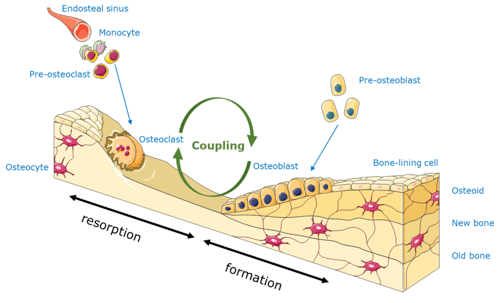
Bone is a very dynamic organ that undergoes constant remodeling throughout life: osteoclasts resorb the bone matrix whereas osteoblasts lay down a new layer of minerals. This process of bone remodeling is necessary to remove old bone matrix and repair stress-induced micro-cracks. This balance of bone resorption and formation is tightly regulated, as an imbalance leads to several bone diseases such as osteoporosis or osteopetrosis.
An important interaction in this well-balanced system is the communication, or also called coupling, between the osteoclasts and osteoblasts. These two different cell types can communicate with each other through direct cell-cell contacts, cytokines or extracellular matrix interactions. Osteoblasts can secrete or express proteins that enhance osteoclastogenesis, such as RANKL or macrophage colony-stimulating factor (M-CSF), as well as proteins that inhibit osteoclast differentiation, such as OPG, also called osteoclastogenesis inhibitory factor (OCIF). On the other hand, osteoclast also affect osteoblast activity by interaction of Semaphorin 4D or EphrinB2. Besides, lots of growth factors, such as TGFβ, BMPs and PDGF are released from the bone matrix upon osteoclast resorption, which enhance the recruitment and differentiation of new osteoblasts (Matsuo and Irie, 2008; Chen et al., 2018).
Title: “Identification of osteoclast-osteoblast coupling factors in humans reveals links between bone and energy metabolism”
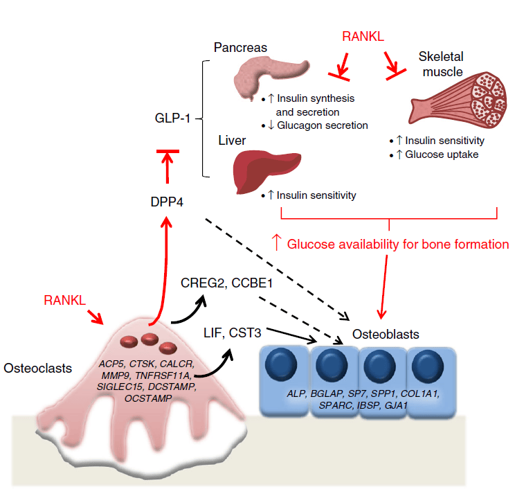
In a recent study, researchers treated postmenopausal women with either placebo or denosumab (DMAb) in order to ablate osteoclast function, and compared the differences in gene expression by RNA-sequencing to find potential osteoclast-derived factors that contribute to osteoclast-osteoblast coupling. After DMAb treatment, they searched for significantly altered secreted factors with the Ingenuity Pathway Analysis and found 55 secreted genes suppressed by the DMAb treatment. To determine which factors are osteoclast or osteoblast derived, they compared enriched populations of osteoblasts and bone marrow-derived osteoclasts by RNA-sequencing and qPCR. From this analysis they found that LIF, CREG2, CTS3, CCBE1, and DPP4 are mostly likely osteoclast-specific factors downregulated by DMAb treatment and potentially involved in the coupling process. As DDP4 is involved in the proteolytic inactivation of glucagon-like peptide-1 (GLP-1), and DDP4 inhibitors are used for the treatment of type 2 diabetes, the researchers wondered if osteoclast-derived DDP4 could influence glucose metabolism. In the postmenopausal women treated with DMAb, they saw an increase in circulating DDP4 levels and GLP1 serum levels, however serum glucose or insulin levels were unchanged, which is expected in patients that don’t have diabetes. Therefore, they examined diabetic patients treated for 1 year with DMAb. In these patients, they did see an improved glycemic control compared to patient on bisphosphonate treatment. The different outcome of both bone resorbing inhibiting drug is probably due to their mechanism of action, where bisphosphonate therapy does not necessarily reduces the number of osteoclasts like DMAb treatment does (Weivoda et al., 2020).
Image from Weivoda M et al. (2020) ‘Identification of osteoclast-osteoblast coupling factors in humans reveals links between bone and energy metabolism’, Nature Communications. Springer US, 11(1), pp. 1-13.
Overall, this study provides outstanding new osteoclast-osteoblast coupling factors in human and observes a potential link between the osteoclast-derived DDP4 and energy metabolism. This information may be of great value in the interpretation of the mechanism of action of bone resorption inhibiting drugs and might guide diabetic-osteoporotic patients towards improved therapeutic treatment.
References:
Chen, Xiao, Wang, Zhongqiu, Duan, Na, Zhu, Guoying, Schwarz, Edward M. and Xie, Chao (2018) ‘Osteoblast–osteoclast interactions’, Connective Tissue Research. Taylor & Francis, 59(2), pp. 99–107.
Matsuo, Koichi and Irie, Naoko (2008) ‘Osteoclast-osteoblast communication’, Archives of Biochemistry and Biophysics, 473(2), pp. 201–209.
Weivoda, Megan M., Chew, Chee Kian, Monroe, David G., Farr, Joshua N., Atkinson, Elizabeth J., Geske, Jennifer R., Eckhardt, Brittany, Thicke, Brianne, Ruan, Ming, Tweed, Amanda J., McCready, Louise K., Rizza, Robert A., Matveyenko, Aleksey, Kassem, Moustapha, Andersen, Thomas Levin, Vella, Adrian, Drake, Matthew T., Clarke, Bart L., Oursler, Merry Jo et al. (2020) ‘Identification of osteoclast-osteoblast coupling factors in humans reveals links between bone and energy metabolism’, Nature Communications. Springer US, 11(1), pp. 1–13.
Currently with the COVID-19 virus chasing through the globe, one can not stop wondering about the secondary effects that might alter people’s life’s after battling the corona infection. As no one exactly knows if this new virus type has long-lasting effects in the hosts body, we can only deduct and speculate from existing data from previous and similar viruses. Here, I will briefly discuss the effect of the immune system on the bone, and the lessons to be learned from the past acute respiratory syndrome (SARS) coronavirus.
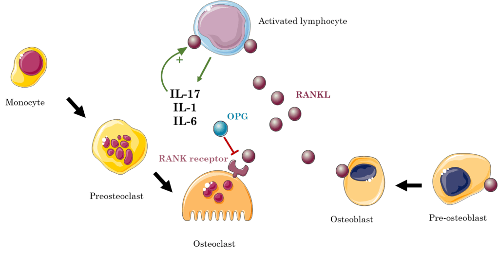
Osteoimmunology is the field which studies the crosstalk and interactions between the skeletal and immune system. An important player in this crosstalk is the RANK/RANKL/OPG signaling pathway. The RANK receptor is expressed by osteoclasts, whereas the RANK ligand (RANKL) is expressed by stromal cells, osteoblasts and immune cells. Additionally, it is found in soluble form within the bone marrow as these cells can secrete RANKL. When the RANKL binds to its receptor, it stimulates osteoclast maturation, activation and survival. To antagonize this signaling pathway, osteoprotegerin (OPG) also has the ability to bind the RANK receptor, thereby preventing RANKL from its binding. Several cytokines, such as interleukin-1 (IL-1), IL-6, IL-17 and tumor necrosis factor α (TNFα) stimulate RANKL expression. Therefore, upon activation of the immune system, activated T and B cells induce osteoclastogenesis, by either directly expressing RANKL, or indirectly by expressing IL-17. Interestingly, in physiological conditions B cells secrete OPG, thus countering the osteogenesis caused by RANK/RANKL signaling. Hence, chronic inflammation might lead to increased bone resorption, giving rise to osteopenia. Due to the fact that viruses activate the immune system, they too might increase bone resorption. Studies have already shown a link between human immunodeficiency virus (HIV) and infection/acquired immunodeficiency syndrome (AIDS) in the formation of osteoporosis, as concomitantly an increase in B cells RANKL expression facilitates bone resorption (Criscitiello et al., 2015; Weitzmann, 2017)
After the outbreak of SARS caused by the SARS coronavirus (SARS-CoV) in 2002, in which patients were treated short-term with high doses of corticosteroids, several reports indicated that recovered SARS patients showed osteonecrosis of hip and knee and reduced hip bone mineral density, which correlated in a dose-correlated manner with the steroid treatment (Griffith et al., 2005; Lau et al., 2005). However, a study by Obitsu and colleagues in 2009 showed that the angiotensin-converting enzyme 2 (ACE2) receptor to which SARS-CoV binds is partially expressed by CD14+ monocytes, a possible precursors of osteoclasts. Furthermore they described that the expression of 3a/X1, an accessory protein of the SARS-CoV, enhanced osteoclastogenesis by increasing RANKL expression in mouse stromal ST2 cells and by enriching the RANKL presence on murine macrophage cell line (Obitsu et al., 2009). This data suggests that upon infection with the virus, more RANKL is produced to bind to their receptor on the osteoclasts, stimulating their maturation and activity, as well as increased susceptibility of monocytes to RANKL, as they generate more receptors.
Notwithstanding, these publications do not anticipate any causal effect between the current coronavirus and bone health, as the previous described bone loss was mainly caused by the steroid treatment, and the direct evidence of SARS-CoV and osteoclastogenesis is limited to in vitro experiments conducted in murine cell lines. Currently, in this pandemic we still don’t know any long-lasting effects caused by the virus, as currently the focus is to prevent its spread and finding a vaccine. If COVID-19 or the immunological response thereon affects bone mass is to be elucidated in the following years.
Criscitiello, Carmen, Viale, Giulia, Gelao, Lucia, Esposito, Angela, De Laurentiis, Michele, De Placido, Sabino, Santangelo, Michele, Goldhirsch, Aron and Curigliano, Giuseppe (2015) ‘Crosstalk between bone niche and immune system: Osteoimmunology signaling as a potential target for cancer treatment’, Cancer Treatment Reviews. Elsevier Ltd, 41(2), pp. 61–68.
Griffith, James Francis, Antonio, Gregory Ernest, Kumta, Shekhar Madhukar, Hui, David Shu Cheong, Wong, Jeffrey Ka Tak, Joynt, Gavin Matthew, Wu, Alan Ka Lun, Cheung, Albert Yu Kiu, Kwok, Hing Chiu, Kai, Ming Chan, Ping, Chung Leung and Ahuja, Anil Tejbhan (2005) ‘Osteonecrosis of hip and knee in patients with severe acute respiratory syndrome treated with steroids’, Radiology, 235(1), pp. 168–175.
Lau, E. M. C., Chan, F. W. K., Hui, D. S. C., Wu, A. K. L. and Leung, P. C. (2005) ‘Reduced bone mineral density in male Severe Acute Respiratory Syndrome (SARS) patients in Hong Kong’, Bone, 37(3), pp. 420–424.
Obitsu, Saemi, Ahmed, Nursarat, Nishitsuji, Hironori, Hasegawa, Atsuhiko, Nakahama, Ken ichi, Morita, Ikuo, Nishigaki, Kazuo, Hayashi, Takaya, Masuda, Takao and Kannagi, Mari (2009) ‘Potential enhancement of osteoclastogenesis by severe acute respiratory syndrome coronavirus 3a/X1 protein’, Archives of Virology, 154(9), pp. 1457–1464.
Weitzmann, M. Neale (2017) ‘Bone and the Immune System’, Toxicologic Pathology, 45(7), pp. 911–924.
Skeletal stem cells (SSCs) are defined by their capacity to self-renew and give rise to the different cell lineages (e.g. osteogenic, adipogenic and chondrogenic lineage), thereby contributing to bone homeostasis and fracture repair. However, the origin and identity of these cells remain largely unknown, as multiple research groups have described various markers characterizing populations of SSCs, which are still very heterogeneous and only partially overlapping with one another (Ambrosi, Longaker and Chan, 2019). Here, they described c-kit, a cell surface marker of several adult stem cells, as a marker to distinguish fetal from post-natal skeletal progenitor cells (He et al., 2020).
Article: “c-kit expression distinguishes fetal from postnatal skeletal progenitors”
Firstly, they investigated if c-kit was expressed by post-natal SSCs. To do so, they generated an inducible reporter mouse line, marking all c-Kit progeny cells with the TdTomato fluorescent reporter while also targeting mature Col1a1+ osteoblasts, to find out if these osteoblasts were derived from a possible c-kit+ SSC. They generated the KitMerCreMer; R26tdTomato; Col2.3-GFP mouse model with the use of the MerCreMer Cre-lox system.
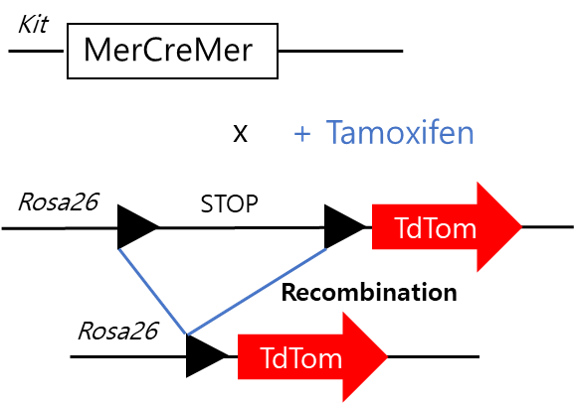
The MerCreMer mouse consists of a Cre recombinase flanked on each side by a mutated murine estrogen receptor (mer) ligand binding domain. Here, this Cre recombinase is under the control of the c-Kit promotor, hence when c-kit is transcribed, so is the MerCreMer. The estrogen binding domain of this Cre recombinase has an affinity for the synthetic estrogen receptor ligands 4-hydroxytamoxifen, or mostly referred to as tamoxifen. Without tamoxifen administration, the Cre recombinase fusion protein is unable to enter the nucleus, and will therefore remain in the cytosol. When tamoxifen is administered, the Cre recombinase enters the nucleus, where it will catalyze the recombination between two loxP sites surrounding, which in this case surrounds a stop codon. This recombination will lead to the deletion of that stop codon, which is followed by a TdTomato sequence. Hence, upon Cre recombination, the TdTomato sequence can be transcribed without problem.
Image adapted from: JH van Berlo et al. Nature 000, 1-5 (2014) doi:10.1038/nature13309
However, no postnatal SSCs expressing c-kit were found. When they administered tamoxifen at postnatal days 1-3, they did not observe any tdTomato expression in growth plate chondrocytes, bone lining osteoblasts or perilipin positive adipocytes, which are all SSC-derived cells. Additionally, by flow cytometry they did not find any Col1a1+ osteoblasts positive for tdTomato expression, confirming that the osteoblasts did not derive from post-natal c-kit positive cells. As during fracture healing the developmental process of endochondral ossification is recapitulated, they performed a femoral fracture at 2 months of age to test whether c-kit is expressed upon re-activation of the post-natal SSCs. At post fracture day 14, they found a lot of newly formed cancellous bone, however none of the osteoblasts were tdTomate positive, indicating they did not derive from c-kit expressing cells. Neither PDGFRα and LepR positive bone marrow cells from the triple negative (hematopoietic) fraction (TN= CD45–Ter119–CD31–), which populations are thought to contain the bone marrow SSCs, were tdTom positive.
Nevertheless, c-kit appeared to mark fetal SSCs. When they administered tamoxifen at E12.5 and E14.5 they did find tdTomato positive growth plate chondrocytes and osteogenic cells in the perichondrium. Furthermore, stromal cells in the primary ossification center were also found tdTom+, indicating the presence of a possible fetal c-kit+ SSC. Interestingly, when they observed these mice at 2-months of age around 20% of TN-PDGFRα+ and TN-LEPR+ were positive for tdTomato, indicating that a portion of these SSC populations were derived from fetal c-kit expressing cells. Additionally, when they performed a colony forming unit (CFU) assay, which represent the bone marrow skeletal stem and progenitor cell (SSPC) population, they found 14% of CFUs to be tdTom+, with the capacity to differentiate in vitro towards the adipo-, chondro- or osteogenic lineage. In vivo, when cultured tdTom+ cells were transplanted subcutaneously to form an ectopic ossicle, these cells had the capacity to form bone with bone marrow and adipocytes, which endorses their stem cell capacities. To characterize the genetic signature of these c-kit derived cells, the researcher performed single-cell RNA sequencing (scRNA-seq) of the TN-PDGFRα+ bone marrow stromal cells from the 2-months old kitMerCreMer; R26tdTomato mice treated with tamoxifen at the embryonic age E12.5/14.5. In this scRNA-seq database they found three main clusters, which were categorized by the expression levels of LepR. However, tdTom+ cells did not show a different expression profile, as the tdTom– and tdTom+ cells were equally distributed along the three clusters.
Furthermore, they tested whether c-kit+ derived cells are functionally important during bone development and SSC cell fate, they generated two different mouse models where important signaling molecules were deleted from c-kit+ cells. Firstly, they deleted mTOR, a signaling molecule involved in many pathways and a regulator for proper skeletogenesis. In this mouse model they observed a decreased bone density due to decreased trabecular number at 2-moths of age, suggesting mTOR signaling participates in the bone forming process of fetal c-kit+ derived osteoblasts. Secondly, they deleted PPARG, a regulator of bone homeostasis and inducer of adipogenesis. In this mouse model however, no differences were observed upon deletion of PPARG of fetal c-kit+ cells. Only a small reduction of perilipin+ cells in the bone marrow was observed, indicating fetal c-kit+ cells give rise to a small portion of adult adipocytes.
Additionally, they investigated the importance of KITL/c-KIT signaling in fetal and post-natal SSCs, they deleted kit-ligand, Kitl, with the use of the Cre-lox system from either only post-natal SSCs, or both fetal and post-natal SSCs, with the Lepr-Cre or Prx1-Cre promotor respectively. They first described the accuracy of their models, as they showed with the use of the TdTomato reporter (either under the LepR or Prx1 promotor) that LepR TdTom+ cells were only found at adult age in the bone marrow, whereas Prx1 TdTom+ cells were found both at fetal and post-natal age. Upon deletion of Kitl in the LepR+ cells they did not observe any differences in microCT analysis of trabecular or cortical bone parameters. In contrast, when they deleted Kitl from Prx1+ cells, they observed increased trabecular bone density, due to increased trabecular number and thickness, indicating that Kitl is a negative regulator of osteogenesis in Prx1+ fetal and post-natal SSCs.
Overall, this study described the expression of c-kit in SSCs during fetal and post-natal life. It shows that fetal SSCs might express different markers than post-natal SSCs, an important aspect to take in consideration while investigating the nature of the SSC.
References:
Ambrosi, Thomas H., Longaker, Michael T. and Chan, Charles K. F. (2019) ‘A Revised Perspective of Skeletal Stem Cell Biology’, Frontiers in Cell and Developmental Biology, 7(September).
He, Di Demi, Tang, Xinyu Thomas, Dong, Wenjie, Cui, Guizhong, Peng, Guangdun, Yin, Xiujuan, Chen, Yujie, Jing, Naihe and Zhou, Bo O. (2020) ‘c-kit expression distinguishes fetal from postnatal skeletal progenitors’, Stem Cell Reports. ElsevierCompany., 14, pp. 1–17.
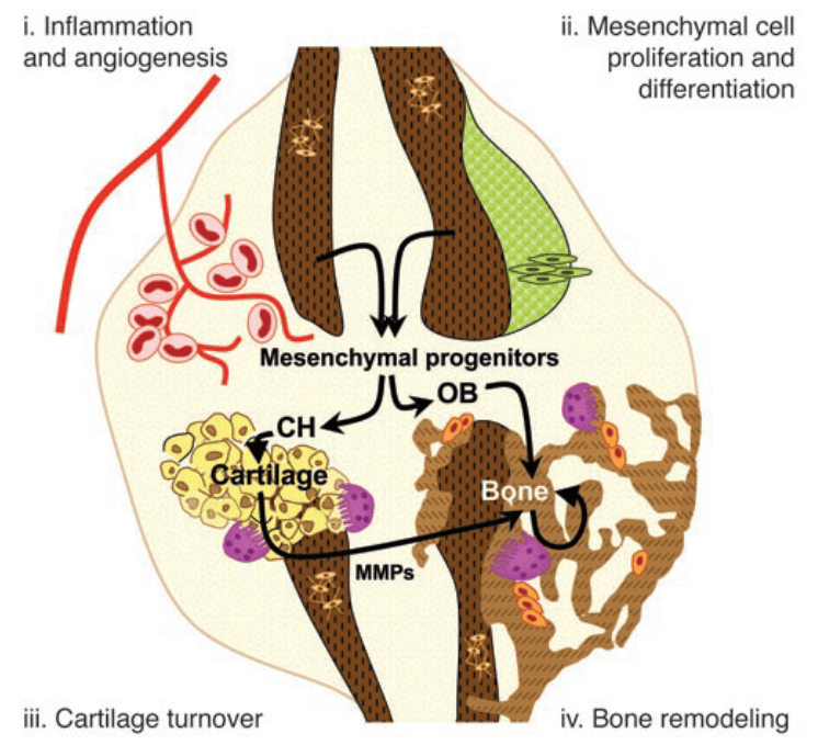
When bone is fractured skeletal stem and progenitor cells activate, proliferate and differentiate into chondrocytes. These chondrocytes will form a cartilaginous template for new bone formation. As blood vessels enter this template, osteoprogenitors come along1, and differentiate into mineralizing osteoblasts. Now, that the template is mineralized, bone-resorbing osteoclasts mingle and reshape the template until the bone is perfectly shaped as before.
Image from Maes, C et al. (2006). Placental growth factor mediates mesenchymal cell development , cartilage turnover , and bone remodeling during fracture repair. Jci 116, 16–182
Understanding the process of fracture healing is of great importance, as 5-10 % of all fractures cannot heal properly. When this happens it severely impacts a person’s life quality, and increases the probability on mortality. As the process of bone formation after a fracture all starts with the skeletal stem cells that divide and give rise to the chondrocytes and osteoblasts, many researchers in the bone field are interested in how this activation process works, and if we could influence it, in order to repair non-healing fractures more easily. Researcher from KU Leuven and Harvard recently discovered how the inflow of nutrients affects the differentiation path the skeletal progenitor cells.3
Article: “Lipid availability determines fate of skeletal progenitor cells via SOX9”
In their paper, they described an increase in chondrogenesis when blood vessels are unable to enter the fractured region. Seven days after the fracture they noticed that blood vessels from the muscles were tightly interwoven with the fracture site and wondered how this interaction was established. Firstly, they examined if the periosteum cells, the cells laying the outer bone surface, contributed to the blood vessel formation. Although they observed that the progenitor cells in the periosteum did not give directly rise to blood vessels, when they removed the periosteum layer, the infiltration of blood vessels was inhibited, therefore suggesting that the periosteum actively promotes the ingrowth of blood vessels. Secondly, they wanted to study the effect on bone repair when blood vessels are absent. Therefore they inserted a filter, wherethrough blood vessels are unable to enter, between the site of injury and the muscle. This resulted in the increase of chondrocytes and reduced bone at the site of injury, together with the increased expression of Sox9, the main transcription factor to start chondrogenic differentiation.
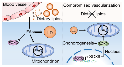
As blood vessels supply the tissue with different types of nutrients, the researchers wanted to know which nutrients were crucial. By depriving cells in vitro with specific nutrients, such as oxygen, glucose and serum, they observed most pronounced effects on the Sox9 levels with the deprivation of serum, a main source for lipids. Moreover, addition of fatty acids to an ex vivo serum deprived culture restored the increased expression of Sox9, and in vivo injection of extra fatty acids into a fracture reduced the amount of chondrocytes present. This suggested that the lipids supplied by blood vessels during fracture healing determine chondrogenic differentiation of the progenitor cells.
Now, which cells need lipids in order to function normally? To answer this question, the researchers looked at the metabolism of osteoblasts and chondrocytes. By using single-cell RNA sequencing of mouse long bones, they found that chondrocytes highly rely on glucose metabolism, whereas osteoblasts use glucose and lipids to obtain their energy. This was confirmed by injecting fluorescent labeled fatty acid and glucose analogues. In this experiment they saw that whereas the glucose was taken up by both cell types, only the osteoblasts consumed the lipids. Therefore, lipids present or not, chondrocytes will survive and thrive anyhow.
Then they looked how the lipid scarcity affects the skeletal stem and progenitor cells. They saw that when skeletal progenitor cells were deprived from lipids, they first responded with the translocation of lipid droplets to the mitochondria, where fatty acid oxidation takes place. This way the skeletal progenitor cells still could maintain themselves within the first hours of lipid scarcity. Afterwards, levels of Sox9 rose concomitantly with the lipid deprivation, indicating a correlation. Therefore, the researchers deleted the Sox9 gene and looked at the response on the lipid metabolism. They saw that cell lacking Sox9 deprived from serum, the main source of lipids, continued lipid acid oxidation, whereas normal cells switched from lipid to glucose metabolism. On the contrary, overexpression of Sox9 reduced lipid consumption, which indicates that Sox9 is a metabolic regulator by suppressing lipid metabolism.
So how does the presence of lipids affects Sox9 levels, which in turn affects lipid metabolism? Upon serum deprivation, the researchers saw an enrichment of the FOXO/forkhead motif coinciding with localization of FOXO1 and FOXO3a protein to the nucleus, where it is transcriptionally active. Inhibition of FOXO signaling seemed to block the increase of Sox9 levels upon lipid deprivation, which indicates FOXO directly affects Sox9 expression during lipid deprivation.
Image taken from Gastel, Nick Van, Stegen, Steve, Eelen, Guy, Schoors, Sandra, Baryawno, Ninib, Przybylski, Dariusz, Depypere, Maarten, Stiers, Pieter-jan, Lambrechts, Dennis, Looveren, Riet Van, et al. (2020). Lipid availability determines fate of skeletal progenitor cells via SOX9. Nature 579, 111–117
These results are of great importance, as they have potential clinical applications in the field of regenerative medicine, as altering the supply of nutrients or manipulating cellular metabolism might drive the progenitor cells into a desired differentiation pathway.
For more details about the experimental work:
Gastel, Nick Van, Stegen, Steve, Eelen, Guy, Schoors, Sandra, Baryawno, Ninib, Przybylski, Dariusz, Depypere, Maarten, Stiers, Pieter-jan, Lambrechts, Dennis, Looveren, Riet Van, et al. (2020). Lipid availability determines fate of skeletal progenitor cells via SOX9. Nature 579, 111–117
References:
1. Maes, Christa, Kobayashi, Tatsuya, Selig, Martin K., Torrekens, Sophie, Roth, Sanford I., Mackem, Susan, Carmeliet, Geert & Kronenberg, Henry M. (2010). Osteoblast Precursors, but Not Mature Osteoblasts, Move into Developing and Fractured Bones along with Invading Blood Vessels. Dev. Cell19, 329–344
2. Maes, Christa, Coenegrachts, Lieve, Stockmans, Ingrid, Daci, Evis, Luttun, Aernout, Petryk, Anna, Gopalakrishnan, Rajaram, Moermans, Karen, Smets, Nico, Verfaillie, Catherine M., Carmeliet, Peter, Bouillon, Roger & Carmeliet, Geert. (2006). Placental growth factor mediates mesenchymal cell development , cartilage turnover , and bone remodeling during fracture repair. Jci116, 16–18
3. Gastel, Nick Van, Stegen, Steve, Eelen, Guy, Schoors, Sandra, Baryawno, Ninib, Przybylski, Dariusz, Depypere, Maarten, Stiers, Pieter-jan, Lambrechts, Dennis, Looveren, Riet Van, et al. (2020). Lipid availability determines fate of skeletal progenitor cells via SOX9. Nature579, 111–117
My name is Dana Trompet, and I am a PhD student in the lab of Skeletal Cell Biology and Physiology at KU Leuven, Belgium. I am passionate about science and would like to share new intersting ideas within my field of expertise.
In this blog I might take you from the depths of bone biology to more general concepts of cellular biology and stem cell biology.
If you have any suggestions, ideas or comments please let me know!
 Bone Biology & Beyond