Skeletal stem cells (SSCs) are defined by their capacity to self-renew and give rise to the different cell lineages (e.g. osteogenic, adipogenic and chondrogenic lineage), thereby contributing to bone homeostasis and fracture repair. However, the origin and identity of these cells remain largely unknown, as multiple research groups have described various markers characterizing populations of SSCs, which are still very heterogeneous and only partially overlapping with one another (Ambrosi, Longaker and Chan, 2019). Here, they described c-kit, a cell surface marker of several adult stem cells, as a marker to distinguish fetal from post-natal skeletal progenitor cells (He et al., 2020).
Article: “c-kit expression distinguishes fetal from postnatal skeletal progenitors”
Firstly, they investigated if c-kit was expressed by post-natal SSCs. To do so, they generated an inducible reporter mouse line, marking all c-Kit progeny cells with the TdTomato fluorescent reporter while also targeting mature Col1a1+ osteoblasts, to find out if these osteoblasts were derived from a possible c-kit+ SSC. They generated the KitMerCreMer; R26tdTomato; Col2.3-GFP mouse model with the use of the MerCreMer Cre-lox system.
The MerCreMer mouse consists of a Cre recombinase flanked on each side by a mutated murine estrogen receptor (mer) ligand binding domain. Here, this Cre recombinase is under the control of the c-Kit promotor, hence when c-kit is transcribed, so is the MerCreMer. The estrogen binding domain of this Cre recombinase has an affinity for the synthetic estrogen receptor ligands 4-hydroxytamoxifen, or mostly referred to as tamoxifen. Without tamoxifen administration, the Cre recombinase fusion protein is unable to enter the nucleus, and will therefore remain in the cytosol. When tamoxifen is administered, the Cre recombinase enters the nucleus, where it will catalyze the recombination between two loxP sites surrounding, which in this case surrounds a stop codon. This recombination will lead to the deletion of that stop codon, which is followed by a TdTomato sequence. Hence, upon Cre recombination, the TdTomato sequence can be transcribed without problem.
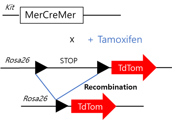
Image adapted from: JH van Berlo et al. Nature 000, 1-5 (2014) doi:10.1038/nature13309
However, no postnatal SSCs expressing c-kit were found. When they administered tamoxifen at postnatal days 1-3, they did not observe any tdTomato expression in growth plate chondrocytes, bone lining osteoblasts or perilipin positive adipocytes, which are all SSC-derived cells. Additionally, by flow cytometry they did not find any Col1a1+ osteoblasts positive for tdTomato expression, confirming that the osteoblasts did not derive from post-natal c-kit positive cells. As during fracture healing the developmental process of endochondral ossification is recapitulated, they performed a femoral fracture at 2 months of age to test whether c-kit is expressed upon re-activation of the post-natal SSCs. At post fracture day 14, they found a lot of newly formed cancellous bone, however none of the osteoblasts were tdTomate positive, indicating they did not derive from c-kit expressing cells. Neither PDGFRα and LepR positive bone marrow cells from the triple negative (hematopoietic) fraction (TN= CD45–Ter119–CD31–), which populations are thought to contain the bone marrow SSCs, were tdTom positive.
Nevertheless, c-kit appeared to mark fetal SSCs. When they administered tamoxifen at E12.5 and E14.5 they did find tdTomato positive growth plate chondrocytes and osteogenic cells in the perichondrium. Furthermore, stromal cells in the primary ossification center were also found tdTom+, indicating the presence of a possible fetal c-kit+ SSC. Interestingly, when they observed these mice at 2-months of age around 20% of TN-PDGFRα+ and TN-LEPR+ were positive for tdTomato, indicating that a portion of these SSC populations were derived from fetal c-kit expressing cells. Additionally, when they performed a colony forming unit (CFU) assay, which represent the bone marrow skeletal stem and progenitor cell (SSPC) population, they found 14% of CFUs to be tdTom+, with the capacity to differentiate in vitro towards the adipo-, chondro- or osteogenic lineage. In vivo, when cultured tdTom+ cells were transplanted subcutaneously to form an ectopic ossicle, these cells had the capacity to form bone with bone marrow and adipocytes, which endorses their stem cell capacities. To characterize the genetic signature of these c-kit derived cells, the researcher performed single-cell RNA sequencing (scRNA-seq) of the TN-PDGFRα+ bone marrow stromal cells from the 2-months old kitMerCreMer; R26tdTomato mice treated with tamoxifen at the embryonic age E12.5/14.5. In this scRNA-seq database they found three main clusters, which were categorized by the expression levels of LepR. However, tdTom+ cells did not show a different expression profile, as the tdTom– and tdTom+ cells were equally distributed along the three clusters.
Furthermore, they tested whether c-kit+ derived cells are functionally important during bone development and SSC cell fate, they generated two different mouse models where important signaling molecules were deleted from c-kit+ cells. Firstly, they deleted mTOR, a signaling molecule involved in many pathways and a regulator for proper skeletogenesis. In this mouse model they observed a decreased bone density due to decreased trabecular number at 2-moths of age, suggesting mTOR signaling participates in the bone forming process of fetal c-kit+ derived osteoblasts. Secondly, they deleted PPARG, a regulator of bone homeostasis and inducer of adipogenesis. In this mouse model however, no differences were observed upon deletion of PPARG of fetal c-kit+ cells. Only a small reduction of perilipin+ cells in the bone marrow was observed, indicating fetal c-kit+ cells give rise to a small portion of adult adipocytes.
Additionally, they investigated the importance of KITL/c-KIT signaling in fetal and post-natal SSCs, they deleted kit-ligand, Kitl, with the use of the Cre-lox system from either only post-natal SSCs, or both fetal and post-natal SSCs, with the Lepr-Cre or Prx1-Cre promotor respectively. They first described the accuracy of their models, as they showed with the use of the TdTomato reporter (either under the LepR or Prx1 promotor) that LepR TdTom+ cells were only found at adult age in the bone marrow, whereas Prx1 TdTom+ cells were found both at fetal and post-natal age. Upon deletion of Kitl in the LepR+ cells they did not observe any differences in microCT analysis of trabecular or cortical bone parameters. In contrast, when they deleted Kitl from Prx1+ cells, they observed increased trabecular bone density, due to increased trabecular number and thickness, indicating that Kitl is a negative regulator of osteogenesis in Prx1+ fetal and post-natal SSCs.
Overall, this study described the expression of c-kit in SSCs during fetal and post-natal life. It shows that fetal SSCs might express different markers than post-natal SSCs, an important aspect to take in consideration while investigating the nature of the SSC.
References:
Ambrosi, Thomas H., Longaker, Michael T. and Chan, Charles K. F. (2019) ‘A Revised Perspective of Skeletal Stem Cell Biology’, Frontiers in Cell and Developmental Biology, 7(September).
He, Di Demi, Tang, Xinyu Thomas, Dong, Wenjie, Cui, Guizhong, Peng, Guangdun, Yin, Xiujuan, Chen, Yujie, Jing, Naihe and Zhou, Bo O. (2020) ‘c-kit expression distinguishes fetal from postnatal skeletal progenitors’, Stem Cell Reports. ElsevierCompany., 14, pp. 1–17.

 Bone Biology & Beyond