Osteoclast secreted proteins link to global energy metabolism

Bone is a very dynamic organ that undergoes constant remodeling throughout life: osteoclasts resorb the bone matrix whereas osteoblasts lay down a new layer of minerals. This process of bone remodeling is necessary to remove old bone matrix and repair stress-induced micro-cracks. This balance of bone resorption and formation is tightly regulated, as an imbalance leads to several bone diseases such as osteoporosis or osteopetrosis.
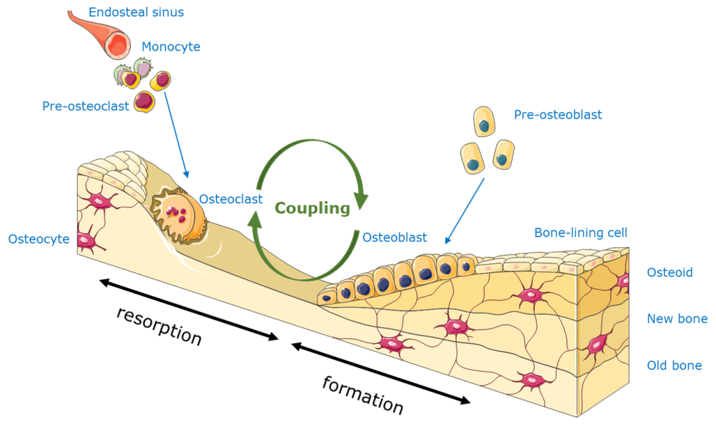
An important interaction in this well-balanced system is the communication, or also called coupling, between the osteoclasts and osteoblasts. These two different cell types can communicate with each other through direct cell-cell contacts, cytokines or extracellular matrix interactions. Osteoblasts can secrete or express proteins that enhance osteoclastogenesis, such as RANKL or macrophage colony-stimulating factor (M-CSF), as well as proteins that inhibit osteoclast differentiation, such as OPG, also called osteoclastogenesis inhibitory factor (OCIF). On the other hand, osteoclast also affect osteoblast activity by interaction of Semaphorin 4D or EphrinB2. Besides, lots of growth factors, such as TGFβ, BMPs and PDGF are released from the bone matrix upon osteoclast resorption, which enhance the recruitment and differentiation of new osteoblasts (Matsuo and Irie, 2008; Chen et al., 2018).

Title: “Identification of osteoclast-osteoblast coupling factors in humans reveals links between bone and energy metabolism”

In a recent study, researchers treated postmenopausal women with either placebo or denosumab (DMAb) in order to ablate osteoclast function, and compared the differences in gene expression by RNA-sequencing to find potential osteoclast-derived factors that contribute to osteoclast-osteoblast coupling. After DMAb treatment, they searched for significantly altered secreted factors with the Ingenuity Pathway Analysis and found 55 secreted genes suppressed by the DMAb treatment. To determine which factors are osteoclast or osteoblast derived, they compared enriched populations of osteoblasts and bone marrow-derived osteoclasts by RNA-sequencing and qPCR. From this analysis they found that LIF, CREG2, CTS3, CCBE1, and DPP4 are mostly likely osteoclast-specific factors downregulated by DMAb treatment and potentially involved in the coupling process. As DDP4 is involved in the proteolytic inactivation of glucagon-like peptide-1 (GLP-1), and DDP4 inhibitors are used for the treatment of type 2 diabetes, the researchers wondered if osteoclast-derived DDP4 could influence glucose metabolism. In the postmenopausal women treated with DMAb, they saw an increase in circulating DDP4 levels and GLP1 serum levels, however serum glucose or insulin levels were unchanged, which is expected in patients that don’t have diabetes.  Therefore, they examined diabetic patients treated for 1 year with DMAb. In these patients, they did see an improved glycemic control compared to patient on bisphosphonate treatment. The different outcome of both bone resorbing inhibiting drug is probably due to their mechanism of action, where bisphosphonate therapy does not necessarily reduces the number of osteoclasts like DMAb treatment does (Weivoda et al., 2020).
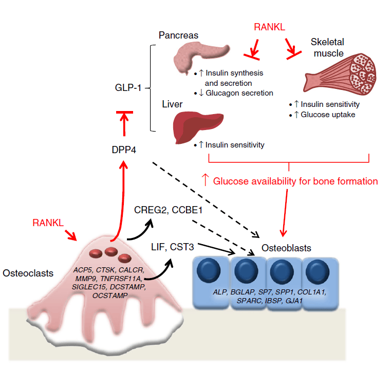
Image from Weivoda M et al. (2020) ‘Identification of osteoclast-osteoblast coupling factors in humans reveals links between bone and energy metabolism’, Nature Communications. Springer US, 11(1), pp. 1-13.

Overall, this study provides outstanding new osteoclast-osteoblast coupling factors in human and observes a potential link between the osteoclast-derived DDP4 and energy metabolism. This information may be of great value in the interpretation of the mechanism of action of bone resorption inhibiting drugs and might guide diabetic-osteoporotic patients towards improved therapeutic treatment.


References:

Chen, Xiao, Wang, Zhongqiu, Duan, Na, Zhu, Guoying, Schwarz, Edward M. and Xie, Chao (2018) ‘Osteoblast–osteoclast interactions’, Connective Tissue Research. Taylor & Francis, 59(2), pp. 99–107.

Matsuo, Koichi and Irie, Naoko (2008) ‘Osteoclast-osteoblast communication’, Archives of Biochemistry and Biophysics, 473(2), pp. 201–209.

Weivoda, Megan M., Chew, Chee Kian, Monroe, David G., Farr, Joshua N., Atkinson, Elizabeth J., Geske, Jennifer R., Eckhardt, Brittany, Thicke, Brianne, Ruan, Ming, Tweed, Amanda J., McCready, Louise K., Rizza, Robert A., Matveyenko, Aleksey, Kassem, Moustapha, Andersen, Thomas Levin, Vella, Adrian, Drake, Matthew T., Clarke, Bart L., Oursler, Merry Jo et al. (2020) ‘Identification of osteoclast-osteoblast coupling factors in humans reveals links between bone and energy metabolism’, Nature Communications. Springer US, 11(1), pp. 1–13.
Could COVID-19 affect the bone?

Currently with the COVID-19 virus chasing through the globe, one can not stop wondering about the secondary effects that might alter people’s life’s after battling the corona infection. As no one exactly knows if this new virus type has long-lasting effects in the hosts body, we can only deduct and speculate from existing data from previous and similar viruses. Here, I will briefly discuss the effect of the immune system on the bone, and the lessons to be learned from the past acute respiratory syndrome (SARS) coronavirus.

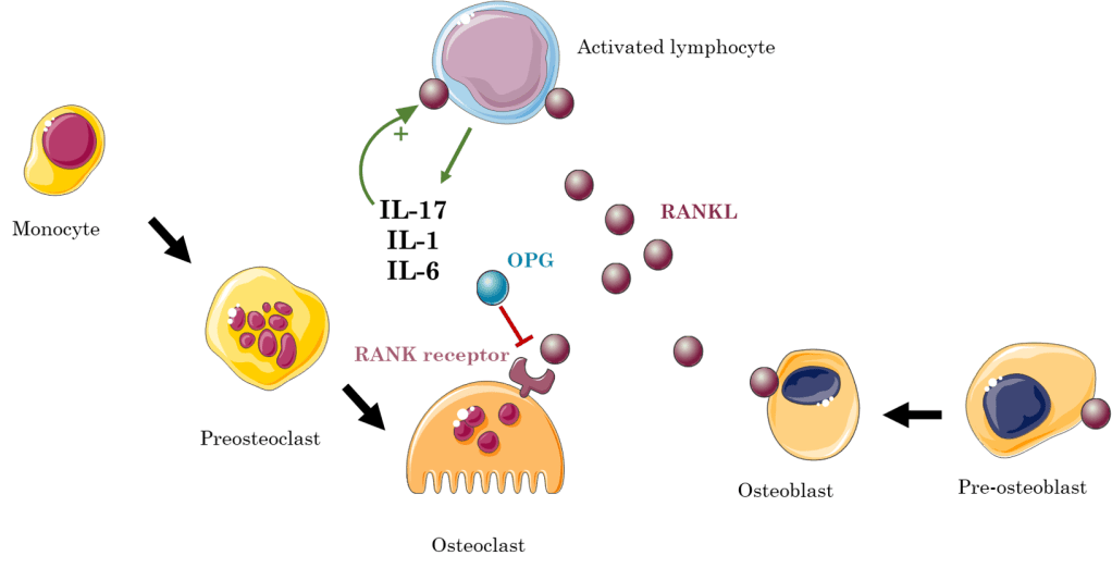
Osteoimmunology is the field which studies the crosstalk and interactions between the skeletal and immune system. An important player in this crosstalk is the RANK/RANKL/OPG signaling pathway. The RANK receptor is expressed by osteoclasts, whereas the RANK ligand (RANKL) is expressed by stromal cells, osteoblasts and immune cells. Additionally, it is found in soluble form within the bone marrow as these cells can secrete RANKL. When the RANKL binds to its receptor, it stimulates osteoclast maturation, activation and survival. To antagonize this signaling pathway, osteoprotegerin (OPG) also has the ability to bind the RANK receptor, thereby preventing RANKL from its binding. Several cytokines, such as interleukin-1 (IL-1), IL-6, IL-17 and tumor necrosis factor α (TNFα) stimulate RANKL expression. Therefore, upon activation of the immune system, activated T and B cells induce osteoclastogenesis, by either directly expressing RANKL, or indirectly by expressing IL-17. Interestingly, in physiological conditions B cells secrete OPG, thus countering the osteogenesis caused by RANK/RANKL signaling. Hence, chronic inflammation might lead to increased bone resorption, giving rise to osteopenia. Due to the fact that viruses activate the immune system, they too might increase bone resorption. Studies have already shown a link between human immunodeficiency virus (HIV) and infection/acquired immunodeficiency syndrome (AIDS) in the formation of osteoporosis, as concomitantly an increase in B cells RANKL expression facilitates bone resorption (Criscitiello et al., 2015; Weitzmann, 2017)

After the outbreak of SARS caused by the SARS coronavirus (SARS-CoV) in 2002, in which patients were treated short-term with high doses of corticosteroids, several reports indicated that recovered SARS patients showed osteonecrosis of hip and knee and reduced hip bone mineral density, which correlated in a dose-correlated manner with the steroid treatment (Griffith et al., 2005; Lau et al., 2005). However, a study by Obitsu and colleagues in 2009 showed that the angiotensin-converting enzyme 2 (ACE2) receptor to which SARS-CoV binds is partially expressed by CD14+ monocytes, a possible precursors of osteoclasts. Furthermore they described that the expression of 3a/X1, an accessory protein of the SARS-CoV, enhanced osteoclastogenesis by increasing RANKL expression in mouse stromal ST2 cells and by enriching the RANKL presence on murine macrophage cell line (Obitsu et al., 2009). This data suggests that upon infection with the virus, more RANKL is produced to bind to their receptor on the osteoclasts, stimulating their maturation and activity, as well as increased susceptibility of monocytes to RANKL, as they generate more receptors.

Notwithstanding, these publications do not anticipate any causal effect between the current coronavirus and bone health, as the previous described bone loss was mainly caused by the steroid treatment, and the direct evidence of SARS-CoV and osteoclastogenesis is limited to in vitro experiments conducted in murine cell lines. Currently, in this pandemic we still don’t know any long-lasting effects caused by the virus, as currently the focus is to prevent its spread and finding a vaccine. If COVID-19 or the immunological response thereon affects bone mass is to be elucidated in the following years.

#COVID-19, #coronavirus #bonebiology #SARS #osteoimmunology

References:

Criscitiello, Carmen, Viale, Giulia, Gelao, Lucia, Esposito, Angela, De Laurentiis, Michele, De Placido, Sabino, Santangelo, Michele, Goldhirsch, Aron and Curigliano, Giuseppe (2015) ‘Crosstalk between bone niche and immune system: Osteoimmunology signaling as a potential target for cancer treatment’, Cancer Treatment Reviews. Elsevier Ltd, 41(2), pp. 61–68.

Griffith, James Francis, Antonio, Gregory Ernest, Kumta, Shekhar Madhukar, Hui, David Shu Cheong, Wong, Jeffrey Ka Tak, Joynt, Gavin Matthew, Wu, Alan Ka Lun, Cheung, Albert Yu Kiu, Kwok, Hing Chiu, Kai, Ming Chan, Ping, Chung Leung and Ahuja, Anil Tejbhan (2005) ‘Osteonecrosis of hip and knee in patients with severe acute respiratory syndrome treated with steroids’, Radiology, 235(1), pp. 168–175.

Lau, E. M. C., Chan, F. W. K., Hui, D. S. C., Wu, A. K. L. and Leung, P. C. (2005) ‘Reduced bone mineral density in male Severe Acute Respiratory Syndrome (SARS) patients in Hong Kong’, Bone, 37(3), pp. 420–424.

Obitsu, Saemi, Ahmed, Nursarat, Nishitsuji, Hironori, Hasegawa, Atsuhiko, Nakahama, Ken ichi, Morita, Ikuo, Nishigaki, Kazuo, Hayashi, Takaya, Masuda, Takao and Kannagi, Mari (2009) ‘Potential enhancement of osteoclastogenesis by severe acute respiratory syndrome coronavirus 3a/X1 protein’, Archives of Virology, 154(9), pp. 1457–1464.

Weitzmann, M. Neale (2017) ‘Bone and the Immune System’, Toxicologic Pathology, 45(7), pp. 911–924.

Difference in c-kit expression of fetal vs postnatal SSCs

Skeletal stem cells (SSCs) are defined by their capacity to self-renew and give rise to the different cell lineages (e.g. osteogenic, adipogenic and chondrogenic lineage), thereby contributing to bone homeostasis and fracture repair. However, the origin and identity of these cells remain largely unknown, as multiple research groups have described various markers characterizing populations of SSCs, which are still very heterogeneous and only partially overlapping with one another (Ambrosi, Longaker and Chan, 2019). Here, they described c-kit, a cell surface marker of several adult stem cells, as a marker to distinguish fetal from post-natal skeletal progenitor cells (He et al., 2020).

Article: “c-kit expression distinguishes fetal from postnatal skeletal progenitors”

Firstly, they investigated if c-kit was expressed by post-natal SSCs. To do so, they generated an inducible reporter mouse line, marking all c-Kit progeny cells with the TdTomato fluorescent reporter while also targeting mature Col1a1+ osteoblasts, to find out if these osteoblasts were derived from a possible c-kit+ SSC. They generated the KitMerCreMer; R26tdTomato; Col2.3-GFP mouse model with the use of the MerCreMer Cre-lox system.

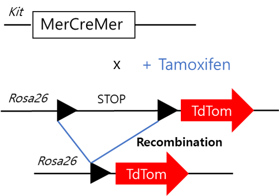
The MerCreMer mouse consists of a Cre recombinase flanked on each side by a mutated murine estrogen receptor (mer) ligand binding domain. Here, this Cre recombinase is under the control of the c-Kit promotor, hence when c-kit is transcribed, so is the MerCreMer. The estrogen binding domain of this Cre recombinase has an affinity for the synthetic estrogen receptor ligands 4-hydroxytamoxifen, or mostly referred to as tamoxifen. Without tamoxifen administration, the Cre recombinase fusion protein is unable to enter the nucleus, and will therefore remain in the cytosol. When tamoxifen is administered, the Cre recombinase enters the nucleus, where it will catalyze the recombination between two loxP sites surrounding, which in this case surrounds a stop codon. This recombination will lead to the deletion of that stop codon, which is followed by a TdTomato sequence. Hence, upon Cre recombination, the TdTomato sequence can be transcribed without problem.

undefined

Image adapted from: JH van Berlo et al. Nature 000, 1-5 (2014) doi:10.1038/nature13309

However, no postnatal SSCs expressing c-kit were found. When they administered tamoxifen at postnatal days 1-3, they did not observe any tdTomato expression in growth plate chondrocytes, bone lining osteoblasts or perilipin positive adipocytes, which are all SSC-derived cells. Additionally, by flow cytometry they did not find any Col1a1+ osteoblasts positive for tdTomato expression, confirming that the osteoblasts did not derive from post-natal c-kit positive cells. As during fracture healing the developmental process of endochondral ossification is recapitulated, they performed a femoral fracture at 2 months of age to test whether c-kit is expressed upon re-activation of the post-natal SSCs. At post fracture day 14, they found a lot of newly formed cancellous bone, however none of the osteoblasts were tdTomate positive, indicating they did not derive from c-kit expressing cells. Neither PDGFRα and LepR positive bone marrow cells from the triple negative (hematopoietic) fraction (TN= CD45Ter119CD31), which populations are thought to contain the bone marrow SSCs, were tdTom positive.

Nevertheless, c-kit appeared to mark fetal SSCs. When they administered tamoxifen at E12.5 and E14.5 they did find tdTomato positive growth plate chondrocytes and osteogenic cells in the perichondrium. Furthermore, stromal cells in the primary ossification center were also found tdTom+, indicating the presence of a possible fetal c-kit+ SSC. Interestingly, when they observed these mice at 2-months of age around 20% of TN-PDGFRα+ and TN-LEPR+ were positive for tdTomato, indicating that a portion of these SSC populations were derived from fetal c-kit expressing cells. Additionally, when they performed a colony forming unit (CFU) assay, which represent the bone marrow skeletal stem and progenitor cell (SSPC) population, they found 14% of CFUs to be tdTom+, with the capacity to differentiate in vitro towards the adipo-, chondro- or osteogenic lineage. In vivo, when cultured tdTom+ cells were transplanted subcutaneously to form an ectopic ossicle, these cells had the capacity to form bone with bone marrow and adipocytes, which endorses their stem cell capacities. To characterize the genetic signature of these c-kit derived cells, the researcher performed single-cell RNA sequencing (scRNA-seq) of the TN-PDGFRα+ bone marrow stromal cells from the 2-months old kitMerCreMer; R26tdTomato mice treated with tamoxifen at the embryonic age E12.5/14.5. In this scRNA-seq database they found three main clusters, which were categorized by the expression levels of LepR. However, tdTom+ cells did not show a different expression profile, as the tdTom and tdTom+ cells were equally distributed along the three clusters.

Furthermore, they tested whether c-kit+ derived cells are functionally important during bone development and SSC cell fate, they generated two different mouse models where important signaling molecules were deleted from c-kit+ cells. Firstly, they deleted mTOR, a signaling molecule involved in many pathways and a regulator for proper skeletogenesis. In this mouse model they observed a decreased bone density due to decreased trabecular number at 2-moths of age, suggesting mTOR signaling participates in the bone forming process of fetal c-kit+ derived osteoblasts. Secondly, they deleted PPARG, a regulator of bone homeostasis and inducer of adipogenesis. In this mouse model however, no differences were observed upon deletion of PPARG of fetal c-kit+ cells. Only a small reduction of perilipin+ cells in the bone marrow was observed, indicating fetal c-kit+ cells give rise to a small portion of adult adipocytes.

Additionally, they investigated the importance of KITL/c-KIT signaling in fetal and post-natal SSCs, they deleted kit-ligand, Kitl, with the use of the Cre-lox system from either only post-natal SSCs, or both fetal and post-natal SSCs, with the Lepr-Cre or Prx1-Cre promotor respectively. They first described the accuracy of their models, as they showed with the use of the TdTomato reporter (either under the LepR or Prx1 promotor) that LepR TdTom+ cells were only found at adult age in the bone marrow, whereas Prx1 TdTom+ cells were found both at fetal and post-natal age. Upon deletion of Kitl in the LepR+ cells they did not observe any differences in microCT analysis of trabecular or cortical bone parameters. In contrast, when they deleted Kitl from Prx1+ cells, they observed increased trabecular bone density, due to increased trabecular number and thickness, indicating that Kitl is a negative regulator of osteogenesis in Prx1+ fetal and post-natal SSCs.

Overall, this study described the expression of c-kit in SSCs during fetal and post-natal life. It shows that fetal SSCs might express different markers than post-natal SSCs, an important aspect to take in consideration while investigating the nature of the SSC.

References:

Ambrosi, Thomas H., Longaker, Michael T. and Chan, Charles K. F. (2019) ‘A Revised Perspective of Skeletal Stem Cell Biology’, Frontiers in Cell and Developmental Biology, 7(September).

He, Di Demi, Tang, Xinyu Thomas, Dong, Wenjie, Cui, Guizhong, Peng, Guangdun, Yin, Xiujuan, Chen, Yujie, Jing, Naihe and Zhou, Bo O. (2020) ‘c-kit expression distinguishes fetal from postnatal skeletal progenitors’, Stem Cell Reports. ElsevierCompany., 14, pp. 1–17.