Bone has long been thought of to be a static organ, providing mechanical support to the body. However, recently several discoveries have shown that bone could also act as an endocrine organ, in which it signals to other organs, influencing glucose metabolism, energy expenditure and male fertility (Karsenty, 2011). More specifically, signaling between the bone and muscle has been described by studying the musculoskeletal system. This system can be seen as one complex organ, which comprises of skeletal bones, muscle, tendons, ligaments, cartilage and joints. These individual parts must be in efficient communication with one another to provide proper locomotion (Brotto and Johnson, 2014).
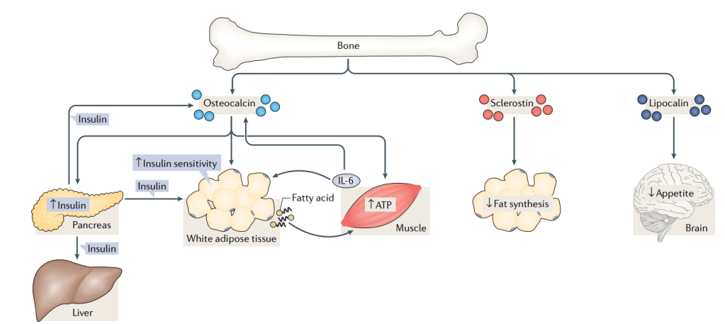
Image derived from (Dirckx et al., 2019); Dirckx, Naomi, Moorer, Megan C., Clemens, Thomas L. and Riddle, Ryan C. (2019) ‘The role of osteoblasts in energy homeostasis’, Nature Reviews Endocrinology. Springer US, 15(11), pp. 651–665.
Osteocalcin is a protein specifically secreted by the osteoblasts during bone formation and is therefore often used as a serum marker for bone formation. Post-translationally it undergoes carboxylation, which increases its affinity to the hydroxyapatite crystals, facilitating the embedding into the bone matrix. Upon bone resorption, which initiates with the formation of an acidic environment by osteoclasts, osteocalcin is decarboxylated and therefore loses its affinity to the bone matrix and promotes the entrance into the circulation (Mizokami, Kawakubo-Yasukochi and Hirata, 2017). In a recent publication, Subrata Chowdhury and colleagues describe the mechanism how osteocalcin secretion by osteoblasts enhances muscle function during endurance exercise (Chowdhury, 2020).
Title: “Muscle-derived interleukin 6 increases exercise capacity by signaling in osteoblasts”
In this work, they studied the interplay between interleukin-6 and osteocalcin upon exercise. Firstly, they observed a correlation between osteocalcin and interleukin-6 (IL-6) serum levels; IL-6 levels increased upon osteocalcin injection in several mammals, and in humans both IL-6 and osteocalcin serum levels increased after exercise. Secondly, they conditionally deleted IL-6 from muscle myofibers by crossing the Hsa-MerCreMer mice with IL-6 floxed mice. In this model, no IL-6 was increased in the serum upon exercise, indicating that IL-6 is mainly muscle-derived. To determine if the changes in serum levels of IL-6 and osteocalcin where directly correlated, they generated heterozygote Ocn+/- IL-6+/- mice. These mice showed similar phenotype compared to the IL-6 conditionally deleted model, indicating a direct connection between the two proteins. An injection of osteocalcin could rescue the reduced exercise capacity, indicating that osteocalcin regulates the IL-6 serum levels.
To uncover an interplay between IL-6 and osteocalcin, the researchers were interested in the cell type present in the bone (osteoblast or osteoclast) that responded to the increased IL-6 levels. To study the interplay between osteoblasts and osteoclasts, they performed a co-culture, where both cell types are cultured within the same medium but aren’t physically touching. This allows the communication with one another by secreted signaling molecules, without any direct contact. They deleted IL-6 receptor in osteoblasts or osteoclasts prior to the co-culture, and studied the effects on osteoclastogenesis. They found that deletion of the IL-6 receptor in osteoblasts, not in osteoclasts, influenced the osteoclastic differentiation, indicating that IL-6 signals through osteoblasts and consequently affects the osteoblast-osteoclast coupling.
To validate this in vitro finding in vivo, they conditionally deleted IL-6 receptor in osteocalcin expressing osteoblasts. Strikingly, these mice showed similar reduction in endurance exercise as they had seen in the conditional model where they deleted muscle-derived IL-6. These mice did not show any increase in osteocalcin upon exercise and the observed phenotype could be partially rescued by administration of osteocalcin, whereas administration of IL-6 did not. Hence, this in vivo model proves that IL-6 signaling through osteoblasts is required for the beneficial osteocalcin derived effects on exercise.
In this study, the researchers demonstrate a new pathway in which bone signals to muscle upon exercise. This research widens the knowledge of the endocrine functions described to bone and makes the field wonder about a more general role of bone and bone-derived cells and their interaction and role within the whole body.
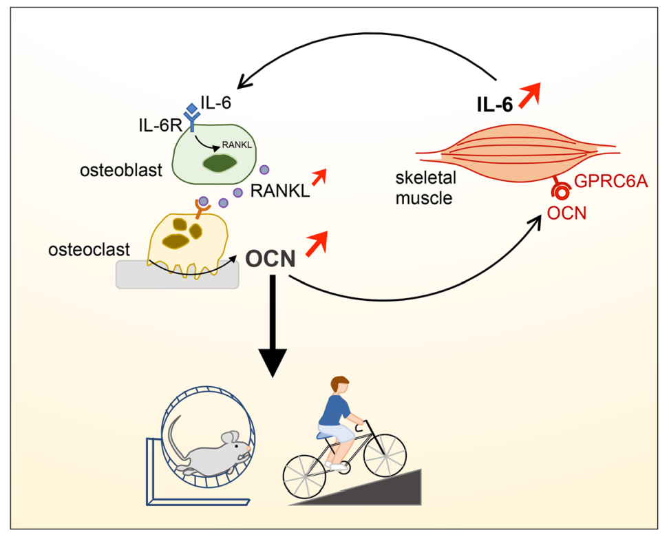
Image derived from Chowdhury S, Schulz L, Palmisano B, et al. Muscle-derived interleukin 6 increases exercise capacity by signaling in osteoblasts [published online ahead of print, 2020 Apr 27]. J Clin Invest. 2020;133572. doi:10.1172/JCI133572
References:
Chowdhury S, Schulz L, Palmisano B, et al. Muscle-derived interleukin 6 increases exercise capacity by signaling in osteoblasts [published online ahead of print, 2020 Apr 27]. J Clin Invest. 2020;133572. doi:10.1172/JCI133572
Brotto, Marco and Johnson, Mark L. (2014) ‘Endocrine crosstalk between muscle and bone’, Current Osteoporosis Reports, 12(2), pp. 135–141.
Dirckx, Naomi, Moorer, Megan C., Clemens, Thomas L. and Riddle, Ryan C. (2019) ‘The role of osteoblasts in energy homeostasis’, Nature Reviews Endocrinology. Springer US, 15(11), pp. 651–665.
Karsenty, Gerard (2011) ‘Bone endocrine regulation of energy metabolism and male reproduction’, Comptes Rendus – Biologies. Academie des sciences, 334(10), pp. 720–724.
Mizokami, Akiko, Kawakubo-Yasukochi, Tomoyo and Hirata, Masato (2017) ‘Osteocalcin and its endocrine functions’, Biochemical Pharmacology, 132, pp. 1–8.
 Bone Biology & Beyond